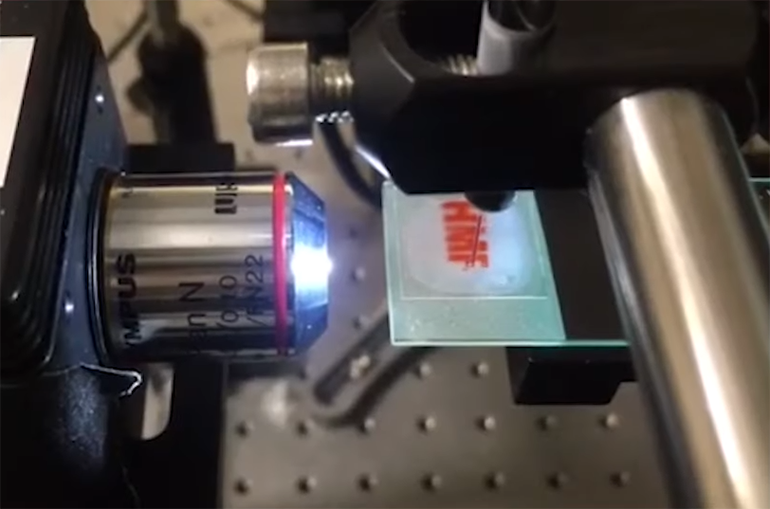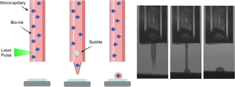
A team at Concordia University in Montreal have developed a technique called Laser-Induced Side Transfer (LIST) that allows for bioprinting of neurons. Low energy laser pulses are directed at a capillary containing a cell-laden bioink, resulting in microbubbles that eject a microjet of the ink onto the substrate below. The technique appears to be fairly gentle on the neurons, and most demonstrated full viability when assessed two days later. The researchers hope that the approach could pave the way for neuron-based printed constructs to assist in drug discovery, and disease modeling.
Bioprinting holds enormous potential in numerous biomedical fields, from drug discovery to regenerative medicine. The technique is similar to 3D printing, but typically involves printing a suspension of cells within a ‘bioink,’ which needs to balance the twin demands of cell viability and appropriate physical and chemical properties for printing.
However, the mechanical, chemical and temperature stresses cells encounter during this process is often too much for them, and maintaining cell viability is a hurdle that any decent bioprinting technique needs to clear. The technology is still in its infancy, but researchers are coming up with ingenious methods to successfully bioprint a variety of cell types for various purposes.
This newest technique uses lasers as the driving force behind the printing, and the researchers have been able to use it to print a cell type that hasn’t been extensively explored in its potential for printing – the dorsal root ganglion (DRG). The research team obtained DRG neurons from the peripheral nervous system of mice, and tested their ability to print them using the LIST system.

Once the neurons were suspended in the bioink, the researchers placed the mixture into a small capillary. They then directed low-energy nanosecond laser pulses onto the capillary, resulting in microbubble formation. The microbubbles eject the ink out of the capillary in a microjet, allowing for precise printing.
The approach appears to lead to high levels of cell survival. When the researchers tested the cells two days after printing, they found that 86% were still alive, suggesting that the technique is gentle enough to create cell constructs using DRG neurons.
The team behind the study is realistic about how far the field of bioprinting has advanced to date, but still sees the potential of the new technique. “In general, people often leap to conclusions when we talk about bioprinting,” said Hamid Orimi, one of the team members, in a press release. “They think that we can now print things like human organs for transplants. While this is a long-term objective, we are very far from that point. But there are still many ways to use this technology.”
See a video about the technology below.
Study in journal Micromachines: Bioprinting of Adult Dorsal Root Ganglion (DRG) Neurons Using Laser-Induced Side Transfer (LIST)
Via: Concordia University
